

|
||||||||||||||||||||||||||||||||||||||||||||||||||||||||||||||||||||||||||||||||||||||||||||||||||||||||||||||||||||||||||||||||||||||||||||||||||||||||||||||||||||||||||||||||||||||||||||||||||||||||||||||||||||||||||||||||||||||||||||||||||||||
Tulane National Primate Research Center Tulane Annual Progress Report 2003-2004 The Tulane National Primate Research Center competes
with the Southwest Foundation for Biomedical Research for the dubious
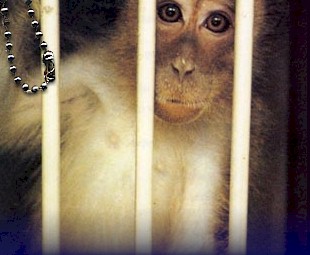
"honor" of imprisoning the largest monkey population among
the eight National Institutes of Health's Regional Primate Research
Centers. Tulane has over 4,500 monkeys of eleven species. Rhesus
macaques form the overwhelming majority with at least 3,500 on hand. Many of the the experiments being funded by the National Institutes of Health conducted at the TNPRC are listed below. Most researchers (e-mail addresses provided) are conducting many studies at any one time. As with other very large primate experimentation sites, it is impossible to keep completely up to date on all the experiments taking place. This snapshot was compiled at the beginning of 2002. BAGBY, GREGORY J. "Ethanol suppression of this response was similar in cells from both control and SIV-infected animals. These results suggest that alcohol might cause a further impairment of host defense in SIV-infected animals and result in an earlier and/or increased incidence of opportunistic infections in these animals....The close similarities between HIV disease in people and SIV infection in rhesus monkeys, together with out initial findings, demonstrate the appropriateness of the rhesus monkey SIV model for the study of the potential impact of alcohol on HIV/AIDS." If the "close similarities between HIV disease in people and SIV infection in rhesus monkeys" had really been demonstrated or were menaingful, the tens of thousands of monkeys killed from experimental SIV infection would have led to some benefit to humans with HIV by now, but it hasn't. Bagby's initial findings are meaningless, as is demonstrated by the relative dearth of other researchers around the country using the same model. BASKIN, GARY B. (a senior veterinarian) <gbask@tpc.tune.edu> writes: "The objectives of this grant are to maintain and propagate a colony of rhesus monkeys that are carriers of globoid cell leukodystrophy (GLD) and to characterize GLD as it occurs in homozygous affected individuals. The long-term goal of this project is to develop a nonhuman primate animal model for gene therapy of inborn errors of metabolism. This colony of rhesus monkeys is the only colony of nonhuman primates in the world in which an inherited lysosomal disorder has been recognized, propagated, and is available for study. No other nonhuman primate model is available in which to test the actual clinical effectiveness of various therapeutic interventions in animals with a genetic disease." Baskin has explained, "An infant rhesus monkey with this disease was diagnosed at TRPRC in 1989. Since then, we intentionally inbred this group of animals and in 1996 observed two additional affected infants." GLD is also called Krabbe's disease. Baskin studiously fails to mention that the disease is well-known in West Highland White Terriers and Cairn Terriers. We wonder whether he believes monkeys are good models of dog diseases. GLD is a rare disease in humans. Baskin also fails to mention that human clinical studies are underway. Clearly, studying and trying to cure humans with the disease is much more likely to lead to a cure for humans than trying to create and study the disease in an alien species. Baskin also fails to mention the suffering that his breeding scheme will create. Symtoms may include loss of previously attained developmental skills, fevers, irritability, myoclonic seizures (sudden shock-like contractions of the limbs), blindness, spasticity (stiffness of the limbs), and paralysis. Prolonged weight loss may occur also. Onset of the disorder in humans is generally between 3 and 6 months of age. BEILKE, MARK A. <mabeilke@tulane.edu> or <idgeno@tulane.edu> "This collaborative project established an animal model of HTLV-I associated disease in nonhuman primates with a primary HTLV-I viral isolate, derived from a human patient with HTLV-I associated tropical spastic paraparesis (TSP). Two of three inoculated rhesus macaques became infected and showed signs of disease. One animal presented with a findings identical to that in the original patient, with muscle atrophy, polymyositis, and arthritis, (manuscript published). The second animal was dually infected with SIV and developed signs suggestive of smoldering HTLV-I induced leukemia in the presence of SIV immunodeficiency disease. Four of five additional HTLV-I inoculated animals are asymptomatic but show evidence of persistent infection by HTLV-I PCR and increased lymphocytosis. The fifth animal is culture and PCR negative with an associated strong humoral response, as determined by western blot analysis." BLANCHARD, JAMES L. (Executive Director) <bubba@tpc.tulane.edu> "The single specific aim of this application is to establish a colony of Indian rhesus monkeys, free of Simian Immunodeficiency Virus (SIV), simian retrovirus (SRV), herpes b virus, and simian t-lymphotrophic virus (STLV 1). This will be accomplished by recruitment of 100 specific pathogen free (SPF) female rhesus monkeys and 15 SPF males in the first year. An additional 50 SPF females and 10 males will be added to the founding colony in each of the subsequent 4 years. We project that by the end of the grant period the colony will have 625 animals." RUDOLF P BOHM, JR <bohm@tpc.tulane.edu> "This study is a continuation of previously reported work to develop a rhesus monkey model of Pneumocystis carinii pneumonia (PCP). This model will be important to study the pathogenesis and novel drug compounds that may be used to treat PCP. Four rhesus monkeys were chosen for this pilot study. Three animals received alternate day dosing of prednisone and cyclophosphamide to induce immunosuppression. The fourth animal was used as a non-immunosuppressed control. Bronchoalveolar lavage (BAL) was performed one week prior to immunosuppression and weekly thereafter. All four animals were inoculated via a bronchoscope with a homogenate from P. carinii frozen lung tissue derived from three SIV infected animals." COGSWELL, FRANK B. <cogswel@tpc.tulane.edu> "Cerebral malaria, a syndrome found in patients infected with Plasmodium falciparum, kills an estimated 2 million children a year. The lack of an animal model has been a barrier to research into supportive therapies and treatment." Frank is profoundly ignorant of the problems associated with health care in the developing world where malaria is common. In these regions, most who contract malaria receive no treatment whatsoever. His claim that the rate of death among children is linked to "the lack of an animal model" is indicative of life in a very lofty ivory tower. Cogswell continues: "We are developing a model of this condition in rhesus monkeys inoculated with P. knowlesi, a simian malaria parasite....We have now selected a population of parasites that consistently adhere to brain endothelium and we will next put these parasites back into a rhesus monkey to produce cerebral malaria. Once this sydrome has been seen in the experimental animals, we will be able to test treatments and supportive therapies that can be taken to endemic areas for use in children afflicted with this condition." Hey Frank, the children in these areas don't have access to the simplest theraputics, like aspirin or clean water. Why not start with the known treatments and cures before trying to invent more therapies that the kids won't get? Does it have anything to do with money? CUSICK, CATHERINE G. <cusick@tulane.edu> "The long term goal of this research is to understand the neural substrates of certain forms of complex visual behavior and other higher cortical functions. To this end, the proposed studies will examine the functional organization of the superior temporal polysensory area (STP) in the superior temporal sulcus (STS) of the rhesus monkey." Cusick plans to place electrodes in the monkeys' brains, shock them to see what happens to their eyes, then inject chemicals into their brains and try to figure out how their brain controls their eyes. DAVISON, BILLIE B. <billie@tpc.tulane.edu> "In studies at TRPRC utilizing simian immunodeficiency virus (SIV)-infected pregnant macaques, chorioamnionitis (CA) [Infection, of the chorionic and amniotic membranes caused by bacteria.] and/or increased inflammatory infiltrates within placental tissues was associated with increased SIV antigen within placental tissues, early fetal demise, and SIV transmission to the infant. During year one (1998-99), this project will develop a primate model of chorioamnionitis by introducing 2 different species and 4 doses of bacteria into the choriodecidual space during pregnancy. During years 2-5, CA will be induced in a group of SIV-infected monkeys and the transmission outcome in this group will be compared to a group of monkeys infected with SIV alone to directly test the hypothesis that increases in inflammatory cell infiltrates within placental tissues increase the rate of transmission of immunodeficiency virus from mother to infant." Besides being simply cruel, Davison appears to be just simple. SIV (simian immunovirus), like HIV, is a disease that damages the immune system. Chorioamnionitis is a bacterial infection. If a bacterial infection has become established, the victim's immune system must be compromised. It can be no surprise that when ill, it is easier to become even more sick when exposed to another pathogen or that, when additionally infected, that the immune system will be further incapacitated. And, his hypothesis is meaningless as well. Few HIV-positive pregnant women can forsee, and thus prevent, acquiring a bacterial infection of the amniotic sack. In another publicly-funded study, this time of malaria's impact on babies, Davison claims, "The information derived from these studies will allow [not might allow, or could allow, but will allow] effective interventions to be designed which will prevent [there's that certainty again] the devastating effects of malaria in pregnant women and their children." Obviously, Davison believes he can see into the future. In this series of monstrous experiments, Davison will: "[D]etermine the effects of parity and prior exposure to Plasmodium [the malaria causing organism] on the course, severity, and outcome of clinical malaria in the mother, fetus, and newborn and to test the hypothesis that macrophage-induced cytokine imbalance causes morphologic and physiologic placental lesions that result in fetal damage, post-natal failure to thrive, and congenital infection." DIDIER, ELIZABETH S. <esdid@tpc.tulane.edu> "Microsporidia cause opportunistic infections in persons with AIDS, organ transplant recipients, children, and travelers. Enterocytozoon bieneusi is the most prevalent microsporidian but attempts to establish a tissue culture system for generating organisms has been unsuccessful. The only nonhuman hosts known for E. bieneusi include pigs and nonhuman primates (eg. Macaca mulatta). Ten SIV-infected rhesus macaques were inoculated orally with E. bieneusi harvested from the stool and duodenal lavage aspirates of human AIDS patients. Spores were detected in stools one week later and continued sporadically for approximately two years or until death of the monkeys, but the inconsistency of spore shedding presently renders this model inadequate for testing antimicrosporidial compounds. However, the monkeys did become infected with E. bieneusi and parasite-associated lesions were identified in the gall bladder, liver, and small intestine. Attempts to infect small animals (eg. gerbils, at hymic mice, immunesuppressed mice) with E. bieneusi also have failed to date." DIDIER, PETER J. (a veterinarian) <pdidier@tpc.tulane.edu> "The objectives of this pilot project are to establish the source of Mycobacterium avium infection in SIV-infected monkeys, characterize immune correlates of disease, and determine the feasibility of vaccination to prevent opportunistic infection of SAIDS animals with M. avium. The incidence of M. avium infection (MAI) in the SAIDS colony has increased from undetectable levels in 1988 to 18-25% in recent years....We have demonstrated that animals infected with SIV and severely immunodeficient can be infected with our pathogenic strain (MavK128) while inoculation with a human strain of M. avium (Serovar 4) fails to infect immunodeficient monkeys." In another study, Peter Didier says: "The objectives of this pilot project are to evaluate changes in ovarian morphology, serum estrogen, bone density, and DNA deletions over time in female rhesus monkeys. Preliminary results indicate that bone strength from young monkeys 3-5 years of age and aged monkeys 22-25 years is different and that breaking strength of femurs decreases substantially with age." HARRISON, RICHARD M. <harrison@tpc.tulane.edu> "This is a continuation of services to other investigators to provide pregnancies of known gestational ages and to provide synchronized menstrual cycles in rhesus monkeys (Macaca mulatta). For timed breeding, the monkeys? cycles are controlled by the daily injection of progesterone (5 mg/day) for10 days. Females are paired with males on days 14 to 21 after the last progesterone injection. Ovulation and conception occurs on day 16 to 18 after the progesterone injections. Success rate, based on viable fetus at 21 days gestation, remains at 60% to 70% for first cycle matings. Fetal development is monitored using ultrasonography. For synchronization of menstrual cycles monkeys are examined twice daily to detect menses. On the 16th days of the cycle daily progesterone injections begin. Injections are continued until all the monkeys to be synchronized have received a minimum of 10 injections. At that point all injections cease. Menses occurs on day 1 or 2 following the last progesterone inj ection. If the investigator wishes to treat the monkeys in the luteal stage of the cycle, progesterone injections can begin again on day 16 - 18 and the monkeys can be maintained in a luteal stage for the study." Few women and few infants (assuming that the monkeys are intended to be models of humans) will be exposed to such a chemical bath. Such unnatural conditions are likely to confound any experimental data and thus render any conclusions meaningless. In another project, Harrison writes: "Reports in the literature suggest that soy-based [infant] formulas may be associated with an increased incidence of goiter and thyroid disease." But, in an experiment that could seem logical only to primate vivisectors and to those who fund them, Harrison placed monkeys in two equal groups. "The experimental group received 8mg/kg body weight of genistein [a chemical found in soy believed to offer protection from some forms of cancer] each Monday through Friday. Dosing continued until day 155 of gestation, at which time a Cesarean section was performed to collect the fetus and placenta. Blood was collected from the maternal peripheral circulation twice weekly during the study period. At delivery blood was collected from the maternal peripheral circulation, the uterine veins, the ovarian veins, and the fetal heart." He discovered more genistein in the blood of the fetuses of mothers who had been given the genistein! LACKNER, ANDREW A. <alackner@tpc.tulane.edu> is continuing to study the monkeys he is killing with SIV. LEVY, LAURA S. <llevy@tulane.edu> "Since 1984, over 1200 necropsies on SIV-infected rhesus and cynomolgus monkeys have been performed at the TRPRC. Lymphoid malignancies [cancer] were detected in a proportion of SIV-infected animals." She seems hopeful of producing more of these cancer infected monkeys. MARTIN, LOUIS N., "SENIOR RESEARCH SCIENTIST," <martin@tpc.tulane.edu> writes: "Regions where HIV infection is prominent frequently overlap malaria endemic regions. However, the impact of coinfection with HIV and malaria is not clear." What is abundantly clear, however, is that in regions of the world where HIV is prominent, almost no medical care is available. Those with HIV in these areas, such as central Africa, will receive no benefit from martin's work. It is doubtful whether anyone will other than Martin and those who are funded through his grant. Martin writes: "We inoculated a monkey with Plasmodium knowlesi. 57 days after SIV inoculation, a time when the SIV infection had already caused a decrease in the percentage of CD4+ cells. Maximum parasitemia (11.6%) occurred 9 days after malaria inoculation and was treated by chloroquine. After treatment the hematocrit recovered steadily through day 26, when malaria re-emerged and a single chloroquine treatment was given. The parasitemia and accompanying decrease in hematocrit rapidly resolved. Malaria re-emerged on day 46 and was again treated. After this third recrudescence and treatment, the parasite did not reappear, so the monkey was reinoculated with malaria. Nine days later the parasite was detected but did not require treatment, remaining a chronic low level infestation. The coinfection with malaria did not affect the progress of SIV infection. The percentage of cells expressing CD4, CD8, CD2, or CD20 did not differ in the malaria-infected monkey compared to other monkeys inoculated with SIV at the same time. No conclusions can be drawn based on 1 monkey, but the feasibility of the SIV/malaria coinfection has been demonstrated. An additional SIV-infected monkey was recently inoculated with malaria 327 days after SIV, a point when the percentage of CD4+ cells had declined markedly. A naive control monkey was inoculated at the same time. It will be interesting to see if the SIV-infected monkey inoculated with malaria at a much later stage of SIV infection will still be able to contain the parasite after 3 treatments, and to see if the ability to control the parasite differs in the control monkey. FUNDING NIH-N01-AI-65310 PUBLICATIONS NONE." MARX, PRESTON A. <pmarx@bellsouth.net> "We ... propose to examine several of the issues concerning sexual transmission in a rhesus macaque model using both SIV and SHIV chimeric viruses. Specifically, we will attempt to define the identity of the first cell(s) infected by SIV in the vaginal and cervical mucosa of macaques....we will also track the kinetics and the pathway of SIV spread from the genital mucosa to the draining (internal iliac) lymph nodes before systemic dissemination. In addition, we will perform a series of experiments to examine directly the issue of selective transmission. Mixtures of SIV variants with known differences in their biological phenotypes will be inoculated both mucosally and intravenously into macaques to determine whether virus with a particular biological prop erty (e.g., macrophage-tropism) will be preferentially selected in animals inoculated mucosally. Similar experiments will also be performed using mixtures of SHIV chimeric viruses...." PHILIPP, MARIO T. <philipp@tpc.tulane.edu> Sometimes, the abstracts found at the CRISP site are written by persons other than those doing the experiments. Such is the case here. It is clear that whomever wrote the abstract is enamored with Dr. Philipp: "Abstract: DESCRIPTION (Adapted from the applicant's abstract): This project, presented by an outstanding group of investigators that includes Drs. M. T. Philipp as P.I. and M. S. Klempner, is designed to complement and expand a human study already in course directed to probe the efficacy of an antibiotic regimen designed to treat chronic Lyme disease." Thirty monkeys will be killed altogether in this study. It remains to be seen: If the study is underway in humans, why use monkeys? If the therapy is successful in humans, why kill monkeys? TRAINA-DORGE, VICKI L. <vicki@tpc.tulane.edu> "A large RSV vaccine study using a Praxis Biologics, formalin-inactivated (FI) RSV vaccine was conducted. Twenty-one seronegative rhesus were divided into seven groups of three monkeys each. Four groups (Gp) received IM injections of the FI-RSV vaccine, ranging from high (250ul) to low (50ul of a 1:25 dilution), in five-fold dilutions, while Gp 5 received the Praxis prepared FI-sham vaccine. An independently prepared, Prince FI-RSV vaccine was used to inoculate Gp6, IM. Finally, Gp 7 was intranasally infected with live RSV. Twenty-one days later, a booster vaccine was administered to the first six groups. Finally, on day 56 post inoculation, a live RSV challenge was administered to all 21 animals....On day 8 post challenge, the animals were necropsied. VEAZEY, RONALD S. <veazey@tpc.tulane.edu> "The objective of this proposal is to carefully define developmental changes in the immunophenotypic composition and function of the systemic and mucosal immune system from neonate to adult in normal and simian immunodeficiency virus (SIV)-infected macaques. In this model, SIV serves as a useful tool to determine which differences between the immature and mature immune system are responsible for the increased susceptibility to SIV." In other words, Ron infects baby monkeys with SIV and then takes notes as he watches them die. WHELTON, PAUL K. <pwhelton@tulane.edu> explains the overall vivisection program at the facility: "The score (sic) of the research projects, although primarily in the area of infectious diseases, covers a range of biomedical disciplines. The proposed AIDS research will include studies of vaccines, the interaction of the IDS virus with other infectious agents, the pathogenesis of disease in the monkey including the transfer of virus form the mother to the fetus, the role of alcohol in the infectious process, and the role of the mucosal immune response. Gene therapy studies will test strategies for correcting globoid cell leukodystrophy, hemophilia B, cystic fibrosis, liver disease, and as a treatment for AIDS. Other infectious disease research will be done on Lyme borreliosis (antibiotic treatment and autoimmunity), malaria (severe cerebral disease and disease during pregnancy)< lymphatic filariasis, and pyelonephritis in diabetics. Other, non-infectious disease research projects will be done on neuropeptides, aging, opioids, and vascular injury with balloon catheters. In addition to the research junction of the Center, this grant will support breeding colonies of macaques. It will also provide support for this Center to serve as a resource for investigators from other institutions who have need to conduct research in non-human primates." [an error occurred while processing this directive] |